What is Intraoperative Monitoring of Nerves?
Intraoperative monitoring, or IOM, is a technique used during surgery to monitor the condition of a patient's nervous system throughout the surgical procedure. Monitoring the condition of the nervous system helps prevent damage to the spinal cord, brain, or nerves. Therefore, the use of intraoperative monitoring reduces the risk of surgery-related nerve damage. IOM is often used during procedures such as spine or brain surgery, where surgical complications may cause a loss of neurological function. Intraoperative monitoring of nerves is also referred to as intraoperative neurophysiological monitoring or intraoperative neuromonitoring.
What are the steps in Intraoperative Monitoring of the Nerves?
Preparation
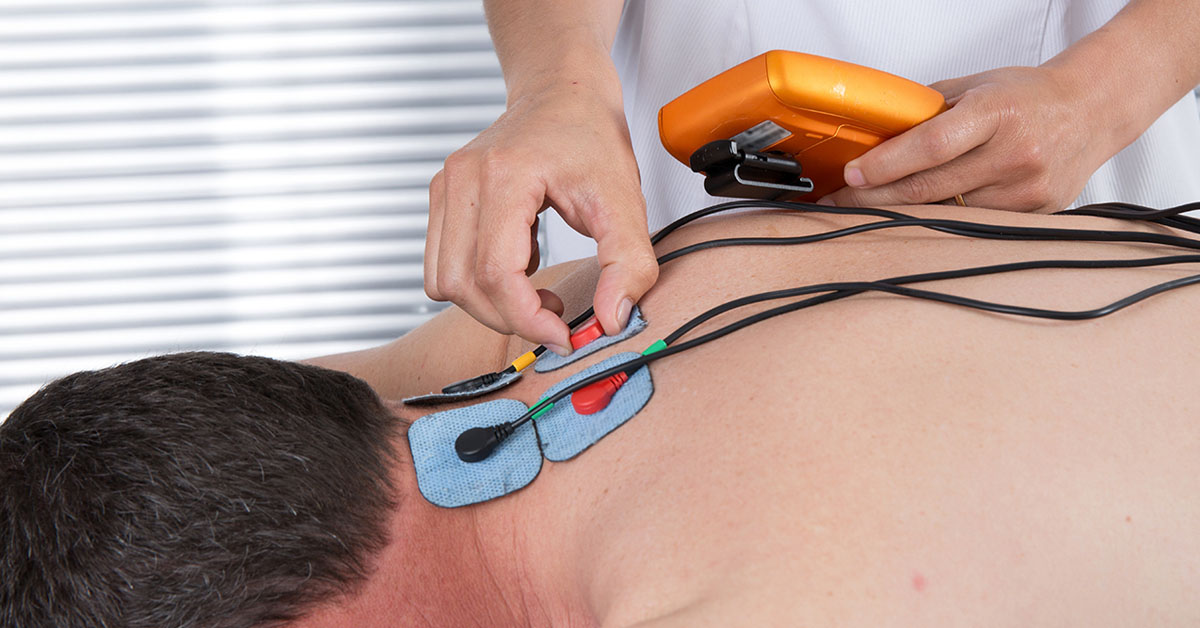
First, electrodes are placed on and/or under the skin at various locations along a nerve pathway. The electrodes are connected to a computer that analyzes information about any nerves being monitored.
Stimulation
Next, an electrode will stimulate the nerve with an electrical impulse. The impulse is brief and contains a very low amount electrical current.
Assessment
Following the stimulation, the nerve's activity is assessed. The other electrodes along the nerve pathway record the time it takes for electrical impulses to travel between them. The speed of the signal is calculated using the distance between electrodes and the time it takes the impulses to travel between them.
Using the Information
The baseline information is compared to that obtain throughout the surgical procedure. If the signal becomes slower and weaker, this reveals a problem with the nerve, like compression. Therefore, changes in signal responses allow a surgeon to quickly identify a problem and take corrective action before nerve damage becomes permanent.
Types of Intraoperative Monitoring
There are several types of intraoperative monitoring that can be utilized during surgery. The main types include:
- Motor Evoked Potentials, or MEPS, allows signals sent from the brain to certain muscle groups to be monitored.
- Somatosensory Evoked Potentials/Dermatome Evoked Potentials, or SSEP/DEPS, allows signals sent to the brain from specific sensory areas to be monitored.
- Electromyography, or EMG, allows signals within certain muscle groups to be monitored during spine surgery. The EMG technique monitors nerves controlling muscle groups during surgery on the part of the spine that controls those muscles. For example:
- During neck, or cervical, surgery: The nerves in the arm muscles are monitored.
- During mid-back, or thoracic, surgery: The nerves in the abdominal muscles are monitored.
- During low back, or lumbar, surgery: The nerves in leg muscles are monitored.