Craniotomy would be needed by patients suffering from conditions within skull, like arteriovenous malformation.
Craniotomy for Arteriovenous Malformations (AVM)
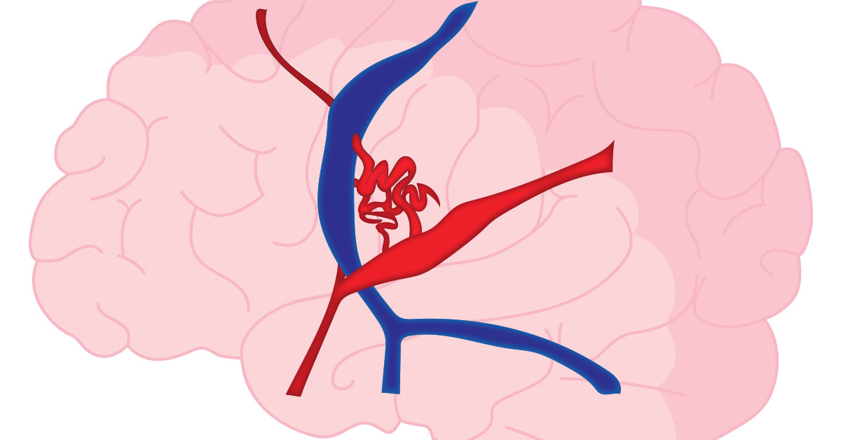
What is an Arteriovenous Malformation?
An arteriovenous malformation, or AVM, is a knot of abnormal blood vessels, like arteries and veins, that is present at birth. They have a high tendency to bleed, most often in young and middle aged adults. AVMs can form in any part of the body, but they are of especially high concern when they form in the brain due to the possibility of bleeding. The size and location of AVMs vary in the brain. AVMs affect less than one percent of the population.
Causes of Arteriovenous Malformations:
Arteries are connected to veins by structures called capillaries. AVMs are formed when arteries connect to veins without the presence of capillaries. AVMs can rupture and damage the tissues of the blood vessels. This results in blood escaping from the vessels and leaking into the brain and surrounding tissues. The flow of blood to the brain is reduced as a result of this bleeding, and the blood clot can damage the surrounding brain.
Symptoms and Diagnosis of Arteriovenous Malformations:
The symptoms depend on the size and location of the arteriovenous malformation. An AVM may not exhibit any symptoms in some cases. Possible symptoms include seizures, headaches and stroke-like symptoms.
The following tests may be used used for diagnosis of arteriovenous malformations: cerebral angiography, magnetic resonance imaging, or MRI, magnetic resonance angiography, or MRA, and computerized tomography, or CT, scans.
Cerebral angiography helps to detect the size and location of arteriovenous malformations. MRI and MRA help produce detailed images of the blood vessels and can highlight AVMs. CT scan can highlight bleeding into the brain.
AVM Treatment Options
The type of treatment is based upon the size and location of the AVM. Treatment options include embolization, radiation therapy, and surgical removal. The advantage of surgical treatment is that a cure is immediate if all the AVM is removed. Disadvantages include risk of bleeding, damage to nearby brain tissue, and stroke to other areas of the brain once removed. AVMs in the brain generally require that a craniotomy be performed in order to treat them.
A craniotomy is a surgical procedure that involves removal of a portion of the skull to access the brain. This surgery is performed by a neurosurgeon while the patient is under anesthesia. A craniotomy can be performed on any part of the skull depending on the location of the brain that needs to be accessed. The portion of the skull that is cut out is called a bone flap.
How is a Craniotomy for AVM Performed?
The following outlines the steps involved in performing a craniotomy:
Before the Procedure
The patient lies on an operating table and is given general anesthesia. Their head is then placed in a device to keep their head in a fixed position during the procedure.
Initial Incision
Some hair may be shaved to clear the incision area. The surgeon makes an incision in the skin. Next, the skin and surrounding muscles are moved aside.
Exposing the Dura
The bone in the exposed part of the skull is cut and the bone flap is lifted, this exposes the covering, called the dura, that protects the brain tissues.
Locating the AVM
The dura is cut and the blood vessel with the AVM is located.
Removing the AVM
The brain tissues around the AVM are separated from the AVM, which is then removed as a single piece.
Ending the Procedure
The surgeon closes the dura and places the bone flap back in its original position. Tiny plates and screws are used to secure the bone flap and keep it in place. Finally, the skin is restored to its original position and sutured.
What Happens After the Craniotomy for AVM is Performed?
After surgery, the patient is moved to an intensive care unit, or ICU, for close monitoring of their vital signs. They may stay on a ventilation and breathing tube for some time. After the procedure, the patients may have a headache or feel nauseated. The patient will remain in the hospital for a few days and receive some short-term rehabilitation.