
Cervical Laminoplasty
Cervical Laminoplasty is a surgical procedure that reshapes or repositions bone in order to relieve stress on the spinal nerves in the cervical section of the spine. Lamina is the roof of bone over the back of spinal cord and plastos means to mold. It is different from a laminectomy because the lamina isn't removed, but repositioned or reshaped.
Who needs Cervical Laminoplasty?
Patients who have cervical stenosis are potential candidates for this surgery. Spinal stenosis occurs when the spinal canal narrows, putting pressure on nerve roots and the spinal cord. The spinal canal may narrow because of the degeneration of the joints and intervertebral discs. Spinal stenosis can also be caused by the formation of bone spurs in the spinal canal or thickening of connecting ligaments.
When the spinal canal becomes narrow, it may start to impinge upon and place pressure on the nerve roots and spinal cord. Symptoms may include:
Feeling of numbness, weakness, and tingling in the neck
Pain in the shoulders, hands or arms
Pain in the neck
Problems in bowel or bladder functions
What are the steps involved in Cervical Laminoplasty?
The steps involved in the Cervical Laminoplasty are as follows.
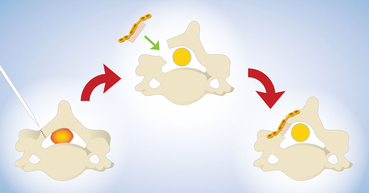
Incision
The patient is given a general anesthesia and is positioned to give the surgeon access to the rear of the neck. The surgeon makes an incision of about three to four inches long. The neck muscles and tissues are dissected to expose the back portion of the vertebrae, called the lamina.
Groove cut on one side
Ones the lamina is exposed, the surgeon cuts a groove on one side of cervical vertebrae to create a hinge.
Cutting of bones on the other side
The surgeon cuts the other side of the cervical vertebrae all the way through. The surgeon then cuts the tips of the spinous processes so that enough room is created for the bones to swing open like a door.
Opening back of vertebrae
The rear of the vertebrae is then opened by the surgeon to take pressure off the nerve roots and spinal cord.
Placing bone wedges
Small wedges made of the bone are placed in the open gap. The bone "door" is then closed by the surgeon. The wedges prevent it from closing completely. Spinal cord and the root nerves rest comfortably.
Closure
The muscles and soft tissues are then put back in their places by the surgeon. The wound is closed by stitching the skin together. The wound is cleaned and the bandage is applied.
What happens after the surgery?
Most of the patients are normally able to get out of bed within one or two hours after the surgery. The surgeon may recommend you to wear a soft neck collar. You will be instructed by the physician to move your neck only very carefully and comfortably.
Returning to work depends on how quickly the patient's body heals and the type of work the patient plans to return to.